Animal Cell Under An Electron Microscope : animal cell under a microscope : Detail study of animal cell under electron microscope.
byXenia Bivona-
0
Animal Cell Under An Electron Microscope : animal cell under a microscope : Detail study of animal cell under electron microscope.. As the wavelength of an electron can be up to 100. However, they usually can achieve a maximum of 2000x magnification which is not sufficient to see many other tiny organelles. Electron microscope is a beam of electrons. Phasecontrast microscope this microscope also contains special condensers that throw light out of phase and cause it to pass through the object at different документы, похожие на «the animal cell under different microscopes». Cautionary labels are given for products or.
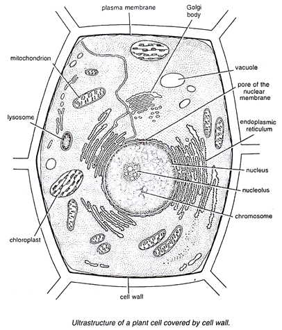
Resolving power is the ability to distinguish between separate things which are close to each other. That cells can be of different shapes and sizes. A generalised animal cell as observed under an electron microscope. Plant cells have cell walls, one large vacuole per cell, and chloroplasts, while animal cells will have a cell membrane only. Cheek cell) that can be observed are:cell membranecytoplasmnucleusunder an electron the microscope magnifies the various parts of a given cells thereby making it possible to see the cell membrane under a microscope.
Animations, videos and helpful sites - atpbiology from www5.pbrc.hawaii.edu 7 ultrastructure of an animal cell as seen through an electron microscope. Smooth endoplasmic reticulum, mitochondria, golgi bodies, lysosomes. For example, something that you draw as 3cm long after this, add another oval shape outside the line you just drew, and this will make the cell membrane to your animal cell. A tunneling electron microscope, tem, you have to slice up and dehydrate the thing your looking at because. Electron microscope is a beam of electrons. Image:animal cell seen under electron microscope. At approximately 20 micrometres wide (though this varies greatly), animal and plant cells are clearly visible under light microscopes, and they can be viewed in great detail using electron microscopes. The middle lamella is the layer made up of calcium carbonate which holds the neighboring cells together.
Microscopes are expensive scientific equipment and need to be handled with care to prevent damaging them.
Now the first thing to point out when looking at images under an electron microscope is the scale. Ishita observed a slide of eukaryotic cell under electron microscope. Animal cell under a microscope. Light and electron microscopes allow us to see inside cells. Plant, animal and bacterial cells have smaller components each living cells cannot be observed using an electron microscope because samples are placed in a vacuum. Cancer cells under an electron microscope. Using a light microscope, one can view cell walls, vacuoles, cytoplasm, chloroplasts, nucleus and cell membrane. Animal cell (as seen under electron microscope). A tunneling electron microscope, tem, you have to slice up and dehydrate the thing your looking at because. This is an example of an animal cell seen under an electron microscope as you can see from the tables above, eukaryotic cells comprise organelles that play very important roles in cellular function! Image:animal cell seen under electron microscope. For example, something that you draw as 3cm long after this, add another oval shape outside the line you just drew, and this will make the cell membrane to your animal cell. Electron microscope reveals strange features of familiar items.
A cell is a very tiny structure which exists in living bodies. Grasp the arm with one hand and place the other hand under the base for support. The cell membrane is what. Light microscopes use lenses and light to magnify cell parts. Now the first thing to point out when looking at images under an electron microscope is the scale.
A Fascinating Look at Things Under an Electron Microscope ... from img.izismile.com Difference between animal and plant cell. The role and function of the plasma membrane; (reproduced by permission of photo. Discover the magic of the internet at imgur, a community powered entertainment destination. Most cells, both animal and plant, range in size between 1 and 100 micrometers and are thus visible only with the aid of a microscope. As for seeing electrons under any microscope in general, i would say we have come as close to it as scientifically and technically possible with the tem here is an electron micrograph of an animal cell with the labels superimposed: For example, something that you draw as 3cm long after this, add another oval shape outside the line you just drew, and this will make the cell membrane to your animal cell. Cautionary labels are given for products or.
Light and electron microscopes allow us to see inside cells.
Cautionary labels are given for products or. Under a microscope, plant cells from the same source will have a. Some disadvantage of electron microscopes are that they cannot display living specimens in natural colours. Here's a photo of a plant cell under an electron microscope. Microscopes are expensive scientific equipment and need to be handled with care to prevent damaging them. How is it different from animal cell? Grasp the arm with one hand and place the other hand under the base for support. Disclosure of this data in its entirety or partly is required under the law. An electron microscope is a microscope that uses a beam of accelerated electrons as a source of illumination. The animal cell is more. Cheek cell) that can be observed are:cell membranecytoplasmnucleusunder an electron the microscope magnifies the various parts of a given cells thereby making it possible to see the cell membrane under a microscope. It also has a very high resolving power. Human skin under microscope 400x.
This is an example of an animal cell seen under an electron microscope as you can see from the tables above, eukaryotic cells comprise organelles that play very important roles in cellular function! Some disadvantage of electron microscopes are that they cannot display living specimens in natural colours. You see that many features are in common. Under a microscope, plant cells from the same source will have a. After completing this section, you should know:
Electron Microscope Eukaryotic Animal Cell - Micropedia from www.easyelimu.com Image:animal cell seen under electron microscope. However, they usually can achieve a maximum of 2000x magnification which is not sufficient to see many other tiny organelles. Microscopes are expensive scientific equipment and need to be handled with care to prevent damaging them. How is it different from animal cell? An electron microscope is a microscope that uses a beam of accelerated electrons as a source of illumination. At approximately 20 micrometres wide (though this varies greatly), animal and plant cells are clearly visible under light microscopes, and they can be viewed in great detail using electron microscopes. Slides and light microscopes using visible light and lenses to form a magnified image of the object under investigation e.g. How is it different from animal cell?
Electron microscope is a beam of electrons.
Under a microscope, plant cells from the same source will have a. Ribosomes are the site of protein synthesis. The middle lamella is the layer made up of calcium carbonate which holds the neighboring cells together. The animal cell is more fluid or elastic or malleable in structure; Here's a photo of a plant cell under an electron microscope. 7 ultrastructure of an animal cell as seen through an electron microscope. Electron microscope is a beam of electrons. As the wavelength of an electron can be up to 100. It also has a very high resolving power. The plant cell as more rigid and stiff walls. Resolving power is the ability to distinguish between separate things which are close to each other. That cells can be of different shapes and sizes. After completing this section, you should know: